.png)

Cardiovascular Imaging includes various tests that take pictures of the heart and its surrounding structures. These allow healthcare workers to diagnose and develop the correct and most effective treatments for different heart conditions.
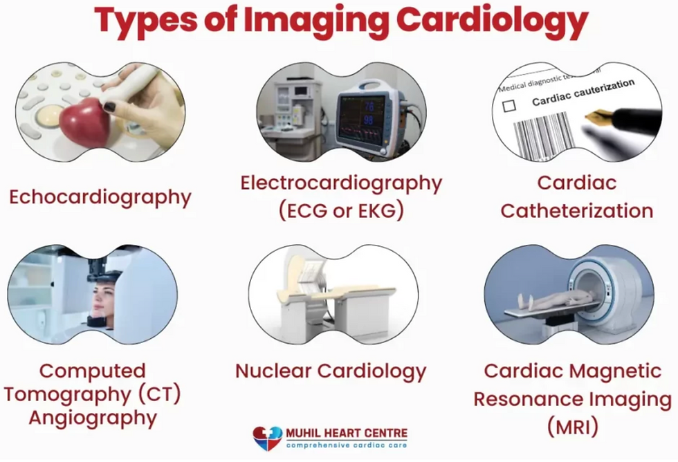
Main types of cardiac Imaging:
Echocardiogram (echo).
Cardiac computed tomography (CT).
Nuclear cardiac stress test.
Single-photon emission computed tomography (SPECT).
Cardiac positron emission tomography (PET).
Coronary angiogram or left heart catheterization ("heart cath").
Each test has unique benefits and uses. Doctors will recommend one or more to their patients depending on the case and their specific health factors. These tests can be used to detect heart problems early, outline the symptoms, and monitor the effectiveness of treatments for conditions like arrhythmia, coronary artery disease, heart attack, heart failure, or heart valve disease. Some symptoms can include chest pain and shortness of breath.
Magnetic Resonance Imaging (MRI)
Cardiac MRI is a noninvasive and painless procedure that generates detailed images of your heart. These images highlight any damage to specific areas and show how well your heart's chambers and valves function.
The heart is 3D and high-resolution images without exposing patients to ionising radiation. It uses massive magnets and radio waves to create high-quality images of your heart on a computer.
Your doctor may recommend a cardiac MRI to diagnose heart problems such as chest pain, shortness of breath, fainting, enlarged heart, thickening of heart muscle, heart failure, heart muscle damage, inflammation, heart valve disease, and infection,
Computed Theart'sgraphy (CT) Scan
Computed tomography (CT) exposes patients to ionizing radiation. Hence, it is only preferred when noninvasive tests are not available and only when it is better suited for low-risk patients. A CT scan involves many X-rays to create high-quality (3D) images of your heart and its surrounding structures. Using multiple X-rays allows us to get images from different angles to create the best quality image to better visualize our heart structure and blood vessels.
Echocardiography
Many doctors prefer them as they provide much information without radiation.
An echocardiogram uses higher-frequency sound waves (ultrasound)to produce accurate images and videos of your heart's chambers, valves, walls, and blood vessels. It can also help identify a valve problem, infection, blood clot or hole in your heart. Furthermore, someone who uses this technology to perform this test is called a sonographer. Additionally, an echocardiogram can help identify various heart problems, such as valve abnormalities, infections, blood clots, or even holes in the heart.
These imaging technologies offer healthcare providers unprecedented insights into cardiac structure and function, allowing for improved procedures, patient care, and better healthcare decisions.
Reference List
Cascino, T. and Shea, M.J. (2023). Cardiac Imaging Tests. [online] MSD Manual Professional Edition. Available at: https://www.msdmanuals.com/en-sg/professional/cardiovascular-disorders/cardiovascular-tests-and-procedures/cardiac-imaging-tests.
Cleveland Clinic. (2022). Cardiac Imaging: Types, Uses and Procedure Details. [online] Cleveland Clinic. Available at: https://my.clevelandclinic.org/health/diagnostics/16836-cardiac-imaging.
Cleveland Clinic. (2022). Echocardiogram: Types and What They Show. [online] Cleveland Clinic. Available at: https://my.clevelandclinic.org/health/diagnostics/16947-echocardiogram.
Cleveland Clinic. (2022). Heart CT Scan: Purpose, Procedure & Risks. [online] Cleveland Clinic. Available at: https://my.clevelandclinic.org/health/diagnostics/16834-cardiac-computed-tomography.
Wikipedia Contributors (2024). Cardiac imaging. [online] Wikipedia. Available at: https://en.wikipedia.org/wiki/Cardiac_imaging#:~:text=Cardiac%20imaging%20refers%20to%20minimally [Accessed 12 Jun. 2024].
Comments